US Pharm. 2010:35:20-23.
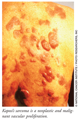
Kaposi's sarcoma (KS) is a multicentric, vascular proliferation considered to be neoplastic and malignant.1 It was first described by the Hungarian dermatologist Moritz Kaposi in 1872.2 Characteristically, KS presents with bluish red or purple discolored skin lesions that are rich in blood vessels and can be associated with widespread visceral involvement.1,3 Among the different forms of KS, the clinical manifestations and course of the disease differ dramatically (TABLE 1).
The classic form of this disease occurs most often in men over 60 years of age of Italian, Jewish, or Eastern European ancestry and is usually not fatal.4 In patients with AIDS, KS develops very quickly and may involve not only the skin, but lungs, gastrointestinal tract, and other organs.3 Several cancers deemed uncommon in the general population are often seen with higher prevalence in transplant recipients--KS being one of them.5 Patients who are immunocompromised experience a particularly virulent, disseminated form of the disease.1 In the elderly, KS may be discovered as an incidental finding.6 Human herpesvirus type 8 has been identified in KS tissue biopsies from practically all patients with the following forms of the disease: classic, endemic, AIDS-associated, and iatrogenic.2,7
Clinical Features and Diagnosis
TABLE 1 provides an overview of the clinical manifestations and prognosis for the different types of KS. Cutaneous Lesions: Most patients have multiple cutaneous lesions that are often bilaterally symmetrical and tend to initially involve the lower extremities; cases have been reported, however, in which only one or no skin lesions have been present.6 These asymptomatic skin lesions are described as purple, pink, or red macules; if fusion occurs, they become plaques with a blue-violet to black hue and eventually form nodules.4 Nodules have the potential to grow rapidly, penetrate soft tissue, and invade bone.1,4 Patients with cutaneous lesions may also present with edema.4
![]()
Mucosal Lesions: The appearance of mucosal lesions is bluish to violaceous (i.e., violet in color) macules, plaques, and tumors.4 While gastrointestinal lesions are usually asymptomatic, they have the potential to bleed; on occasion, the bleeding may be extensive.4
Other Symptoms and Complications: Lesions in the lung may cause cough, shortness of breath, and bloody sputum.3 Pleuropulmonary KS is considered an ominous sign; it usually occurs late in the course of AIDS-associated KS, particularly in patients whose death is directly attributed to KS.2 If lymph nodes are affected, leg swelling may cause pain or infection.3 An aggressive form of endemic KS can quickly spread to the bones.3
Diagnosis: Diagnosis is confirmed by biopsy.3 A CT of the abdomen and chest is performed to evaluate for visceral spread in patients with AIDS or immunosuppression.4 If CT is negative in patients with gastrointestinal or pulmonary symptoms, a gastrointestinal endoscopy or bronchoscopy should be considered.3,4
Characteristics of KS in Seniors
The onset of KS in the elderly is often subtle and, as previously mentioned, with a predisposition to American men of Italian, Jewish, and Eastern European ancestry.4,6 Cutaneous patches and papules are violaceous and present on the lower extremities; lesions of the gastrointestinal tract often require endoscopy for visualization. KS in the elderly is often associated with diabetes mellitus and an increased incidence of
lymphoma.6
Treatment
Lesions may return after treatment.3 Treatment for KS does not improve survival from AIDS itself; prognosis depends on the patient's immune status and viral load.3
Indolent Lesions: Often, no treatment is necessary for indolent (slow growing and causing little pain) lesions; excision, cryotherapy, or electrocoagulation may be used for treatment of one or a few superficial lesions.4 Also useful is intralesional vinblastine (intravenous use only; dose adjusted based on clinical response) or interferon-alfa (e.g., for AIDS-associated KS, interferon-alfa- 2b 30 million units/m2 SC/IM 3 times per week; dosage is decreased by 50% or discontinued if not tolerated).4,8,9 Radiation therapy is utilized for more extensive disease, such as the treatment of multiple lesions and lymph node disease.4
AIDS-Related KS: Antiretrovirals provide the best treatment for AIDS-associated KS, as this condition responds markedly to highly active antiretroviral therapy (HAART).4 In AIDS-associated KS, patients with indolent disease and CD4 counts greater than 150/µL and HIV RNA less than 500 copies/mL can be treated with intravenous interferon-alfa (see above).4 Liposomal doxorubicin (e.g., 20 mg/m2 intravenously every 3 weeks) can be administered to patients with more extensive or visceral disease; paclitaxel (e.g., 100 mg/m2 intravenously every 2 weeks) may be used if doxorubicin fails.4,9 Investigations continue regarding adjunct agents.
Iatrogenic KS: While not always possible, the best treatment response for iatrogenic KS is the discontinuation of immunosuppressants.4 A reduction of KS lesions has been seen in organ transplant patients when the immunosuppressant dosage is reduced; for patients who are unable to tolerate such a reduction, experts recommend local and systemic therapies used in the other types of KS.4
Conclusion
Kaposi's sarcoma is associated with infection by human herpesvirus type 8, which has been identified in KS tissue biopsies in patients with all forms of the disease. Pharmacists should be aware of the clinical manifestations and course of KS and how they differ dramatically depending on the particular form of the disease. KS may also be discovered in senior men as an incidental finding.
Note: Refer to individual drug product labeling for FDA-approved status and individualized dosing guidelines.
REFERENCES
1. Dorland's Pocket Medical Dictionary. 28th ed. Philadelphia, PA: Elsevier Saunders; 2009:743.
2. General Information About Kaposi Sarcoma. National Cancer Institute. U.S. National Institutes of Health [updated September 1, 2009.] www.cancer.gov/cancertopics/pdq/treatment/kaposis/HealthProfessional/page2. Accessed March 15, 2010.
3. Kaposi's Sarcoma. MedlinePlus. National Library of Medicine. National Institutes of Health. Updated September 28, 2008. www.nlm.nih.gov/medlineplus/ency/article/000661.htm. Accessed March 15, 2010.
4. Beers MH, Porter RS, Jones TV, et al. The Merck Manual of Diagnosis and Therapy. 18th ed. Whitehouse Station, NJ: Merck Research Laboratories; 2006:1024-1025.
5. Johnson HJ, Schonder KS. Solid organ transplantations. In: DiPiro JT, Talbert RL, Yee GC, et al, eds. Pharmacotherapy: A Pathophysiologic Approach. 6th ed. New York, NY: McGraw-Hill Inc; 2005:1613-1643.
6. Schwartz RA, Cohen PJ. Kaposi's sarcoma. In: Newcomer VD, Young EM Jr, eds. Geriatric Dermatology: Clinical Diagnosis and Practical Therapy. New York, NY: Igaku-Shoin; 1989:645-652.
7. Samuelson J. Infectious diseases. In: Cotran RS, Kumar V, Collins T, eds. Robbins Pathologic Basis of Disease, 6th ed. Philadelphia, PA: WB Saunders Company; 1999:361.
8. Howland RD, Mycek MJ. Pharmacology. 3rd ed. Philadelphia, PA: Lippincott Williams & Wilkins; 2006:436,471-472,483.
9. Epocrates Rx. Version 9.0. San Mateo, CA: Epocrates, Inc. www.epocrates.com. Accessed March 28, 2010.
10. Schoen FJ, Cotran RS. Blood vessels. In: Cotran RS, Kumar V, Collins T, eds. Robbins Pathologic Basis of Disease, 6th ed. Philadelphia, PA: WB Saunders Company; 1999:535-537.
11. Volberding PA. Hematology and oncology in patients with human immunodeficiency virus infection. In: Goldman L, Ausiello D, eds. Cecil Medicine. 23rd ed. Philadelphia, PA: Saunders Elsevier; 2007:chap416.
To comment on this article, contact rdavidson@uspharmacist.com.


