US Pharm. 2017;42(2):36-39.
ABSTRACT: Venous ulcers, also referred to as venous stasis ulcers (VSUs), are the most common cause of ulcerations that affect the lower extremities. These types of ulcers are experienced by an estimated 1% of the U.S. population. The incidence of VSUs is most prevalent among the elderly population and is also ubiquitous in patients with a medical history of edema in the legs, long-standing varicose veins, or blood clots in either the superficial or the deep veins of the legs. VSUs generally occur in the lower extremities, especially along the medial distal ankle. In general, treatments for VSUs include compression therapy, local wound care and debridement, various types of wound dressings, antibiotics for infected wounds, the use of pharmacologic agents such as pentoxifylline, aspirin, calcium channel blockers, and topical corticosteroids when warranted, as well as surgery and other forms of adjunctive therapy.
Venous ulcers, also referred to as venous stasis ulcers (VSUs), are perceived to be the most common cause of ulcerations affecting the lower extremities and can be severe and debilitating in nature.1-3 VSUs affect an estimated 1% of the U.S. population and are responsible for more than 80% of lower extremity ulcerations.1-3 Statistics also report that an estimated 2.5% of patients admitted to long-term care facilities have VSUs.4
The probability of developing a VSU is directly proportional to increasing age, and its occurrence is also common in patient populations with a history of venous insufficiency.1-5 VSUs occur more frequently in the elderly patient population for several reasons, including calf muscle pump failure, immobility, chronic edema, and obesity.6 Ulceration is precipitated by decreased diffusion of oxygen to the skin and microvasculature damages.4,5 Moreover, the incidence is also greater among paraplegic patients owing to the fact that the calf muscles are immobile.4,5
The healing rates associated with VSUs are often poor, and an estimated 50% of these types of ulcers are still open and unhealed for 9 months or longer.4,5 Therefore, increasing awareness about early recognition, clinical intervention, and patient compliance with therapy is critical to enhancing healing and positive patient outcomes. VSU recurrence rates are also disconcerting, as more than one-third of patients treated for VSUs experience four or more recurrences.4,5
In the United States, VSUs have been estimated to contribute to significant direct and indirect health-care and personal costs including loss of productivity, which accounts for more than 2 million days of absence from work and decreased quality of life. VSUs reportedly incur treatment costs of approximately $3 billion per year.1
While most VSUs are caused by chronic venous insufficiency (CVI), it is imperative that clinicians rule out other underlying causes such as arterial, metabolic, neuropathic, hematologic, infectious, malignant, and inflammatory diseases.6 The differential diagnosis for the VSU may include ischemic ulceration, pyoderma gangrenosum, lymphedema, trauma, neuropathic ulceration, and cellulitis.7 Other possible causes of these types of ulcerations include inflammatory processes, resulting in leukocyte activation, endothelial damage, platelet aggregation, intracellular edema, arterial insufficiency, diabetic neuropathy, prolonged pressure, and chronic conditions such as rheumatoid arthritis, osteomyelitis, and vasculitis.1,4 VSUs are five to seven times more common than the arterial ulcers observed in those with peripheral artery disease.8
VSUs are most commonly seen in patients with a history of leg swelling, varicose veins, or blood clots of the superficial or deep veins of the legs.7-9 Predisposing factors include diabetes mellitus, congestive heart failure, hypertension, and obesity. Diabetic patients with a VSU have a clinical profile and prognosis comparable to those of nondiabetic patients.8,10,11
VSUs occur primarily on the lower extremity over bony prominences, especially along the medial distal ankle and mostly on the inner part of the leg, but the ulcers can occur anywhere on the lower leg and may affect one or both extremities.1-4,7
Causes of VSUs
Certain factors may increase an individual’s risk of developing a VSU. The primary risk factors include older age, previous leg injury, deep venous thrombosis (DVT), and phlebitis.12,13 Examples of other risk factors that may cause CVI that could lead to the development of VSUs include12,13:
- Uncontrolled or undiagnosed diabetes
- History of lower leg edema
- History of DVT
- Uncontrolled or undiagnosed hypertension
- Standing or sitting for long periods
- Obesity
- Family history of VSUs
- Varicose veins
- Leg trauma.
Signs and Symptoms
Patients with VSUs may present with a variety of symptoms depending upon the severity of the ulceration. Patients may present with dull aching or pain in the lower extremity, along with some edema that subsides upon elevation of the affected extremity.7,13,14 The skin may appear erythematous and have a fibrous or eczematous appearance as well.7,13,14 The surrounding skin may present as taut and shiny with a brown or purple pigmentation.7,13 Upon physical examination, the patient may present with ulceration that is large in appearance and is typically shallow, with irregularly shaped edges.2,7,13,14 In many cases, the VSUs are painful, and drainage may also be present.7,13,14 Granulation tissue and fibrin are often present in the ulcer base, and other clinical findings may include lower extremity varicosities; edema; venous dermatitis along with hyperpigmentation and hemosiderosis or hemoglobin deposition in the skin; and lipodermatosclerosis associated with thickening and fibrosis of normal adipose tissue under the skin.2,13,14
Treatment Recommendations
Preventing VSUs is the most important aspect of CVI management; however, patients still develop VSUs, and the goals of therapy include reducing edema, promoting healing of the VSU, and preventing VSU recurrence.14 Failure to treat VSUs may lead to severe complications including infection and/or amputation and increased risk of morbidity and mortality.14 Treatment typically requires on average at least 6 to 12 months of continual therapy to promote healing, but more than 70% of patients experience a recurrence within 5 years.7 It is crucial for clinicians to individualize treatment based on the patient’s overall health, medical conditions, current medications, and psychological status as well as physical ability and willingness to routinely care for the wound.15-18
The Association for the Advancement of Wound Care (AAWC) recommends compression therapy and limb elevation to reduce edema.15 It also recommends cleaning the ulcer with a nonirritating cleanser, debriding nonvital tissue, maintaining a moist wound environment, monitoring for signs of infection, and managing pain and odor.15-18 The most important goal for the treatment of VSUs is long-term edema control.7,15-18 The revised validated VSU guidelines by the AAWC recommend delivery of evidence-based care by qualified professionals via a multidisciplinary team for effective venous ulcer treatment.7 Therefore, it is imperative to get a thorough history and physical examination and consult the proper team members for development of a collaborative treatment strategy.7
The Wound, Ostomy and Continence Nurses Society (WOCN) and the American Society of Plastic Surgeons (ASPS) endorse comparable recommendations, such as ulcer debridement, edema management, infection control, and pain management.17-19 Treatments for VSUs include compression therapy, local wound care (including debridement), wound dressings, topical or systemic antibiotics for infected wounds, other pharmacologic agents, surgery, and adjunctive therapy if warranted.14,16
Compression Therapy
The use of compression therapy with high stiffness index is considered to be the standard of therapy for VSUs. However, patient compliance with compression therapy is sometimes challenging in some populations, particularly in the elderly, because of reduced dexterity, muscle strength, range of motion, and flexibility.6
Until a few years ago, compression bandages were regarded as first-line therapy of VSUs.6,16,20 However, to date medical compression stockings are the first choice of treatment, and they should exert a pressure of at least 20 to 30 mmHg at the ankle to provide efficacious therapy.16,20 Compression therapy helps prevent reflux, decreases release of inflammatory cytokines, and reduces fluid leakage from capillaries, thereby controlling lower extremity edema and VSU recurrence.14,16 Goals of compression therapy are to reduce symptoms, prevent secondary complications, and slow disease progression.14,16 In patients with severe cellulitis, compression therapy is delayed while infection is treated.13,14,16 Contraindications for compression therapy include heart failure, recent DVT, unstable medical status, and risk factors that can cause complications of compression therapy.13,14,16 Additionally, pneumatic compression generally is reserved for patients who cannot tolerate continuous compression.16
Wound Debridement
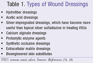
Wound debridement is essential in treating chronic VSUs.13,16 Removing necrotic tissue and bacterial burden through debridement enhances wound healing.13,16 Dressings are used under compression bandages to promote healing, control wound exudate, improve patient comfort, and prevent the wound from adhering to the bandage.14,16 Vacuum-assisted wound-closure therapy can be used with compression bandages.14,16 Examples of the types of available dressings can be found in TABLE 1.14,16
Antibiotics
Both bacterial colonization and infection are common in those with VSUs and contribute to poor wound healing.3,13,16 The use of oral antibiotics is recommended only in cases of suspected wound-bed infection and cellulitis.3,13,16 Parenteral antibiotics are indicated for patients with one or more of the following signs and symptoms associated with infection16:
- Increased erythema of surrounding skin
- Increased pain, local heat, tenderness, and leg swelling
- Quick increase in wound size
- Lymphangitis
- Presence of fever.
Additionally, suspected osteomyelitis requires an evaluation for arterial disease and possible utilization of oral or IV antibiotics to treat the underlying infection, if needed.3,13,16
Adjunctive Therapy
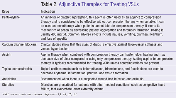
In addition to antibiotics, clinicians may prescribe other pharmacologic agents such as pentoxifylline, calcium channel blockers, aspirin, or topical corticosteroids to treat VSUs (TABLE 2).13,14,16,21 Several other adjunctive therapies are used in the treatment of VSUs including pulsed ultrasound, pulsed electromagnetic fields (PEMFs), and electrical stimulation, which can help facilitate treatment of VSUs that fail to close or show signs of healing despite good conventional wound and compression therapy.13,16,21
Surgery
In some cases, especially in those patients with severe VSUs, surgery may be warranted and may decrease venous reflux, quicken healing, and prevent other episodes of VSUs.13,16 The possible surgical options include ablation of the saphenous vein; interruption of the perforating veins with subfascial endoscopic perforator surgery (SEPS); treatment of iliac vein obstruction with stenting; and removal of incompetent superficial veins with phlebectomy, stripping, sclerotherapy, or laser therapy.13,22,23
The Role of the Pharmacist in Educating Patients About VSUs
Since pharmacists are often perceived as one of the most accessible healthcare professionals, they have a critical role in the treatment and care of patients at risk of, or with, VSUs. This includes effective patient counseling and education, especially with regard to therapy for VSUs, proper use of medications used to treat VSUs, and reminders about preventive measures to decrease reoccurrence of VSUs. Since the incidence of VSUs is common in elderly and diabetic patient populations, pharmacists play a key role in identifying those patients at greater risk of developing VSUs. Pharmacists should also seize every possible opportunity to remind patients about the critical importance of adhering to therapy to enhance therapeutic outcomes and prevent further complications.
Pharmacists can also reinforce patient education with regard to the preventive measures for VSUs such as staying vigilant about one’s overall health and adhering to the goals of therapy. Patients should be reminded of the importance of routine wound care and follow-up with their primary healthcare provider, monitoring the ulcer for signs of infection, and immediately seeking medical attention if the wound shows signs of worsening or not healing. Patients at risk for VSUs should be aware that with early diagnosis and implementation of and adherence to therapy, the condition can be effectively treated and healed to avoid complications.
REFERENCES
1. Agale SV. Chronic leg ulcers: epidemiology, aetiopathogenesis, and management. Ulcers. 2013;2013:413604.
2. de Araujo T, Valencia I, Federman DG, Kirsner RS. Managing the patient with venous ulcers. Ann Intern Med. 2003;138(4):326-334.
3. O’Meara S, Al-Kurdi D, Ovington LG. Antibiotics and antiseptics for venous leg ulcers. Cochrane Database Syst Rev. 2008;(1):CD003557.
4. Gabriel A. Vascular ulcers. Medscape. http://emedicine.medscape.com/article/1298345-overview#a6. Accessed November 1, 2016.
5. Fishman TD. How to manage venous stasis ulcers. Podiatry Today. 2007;20(5). www.podiatrytoday.com/article/7071. Accessed November 1, 2016.
6. Woo KY, LeBlanc K, Livingston. Management of venous stasis ulcers in the older adult. Curr Geri Rep. 2015;4:229-236.
7. Park NJ, Allen L, Guosheng G, Driver VR. Essential insights on treating chronic venous stasis ulcers. Podiatry Today. 2012;25(7). www.podiatrytoday.com/essential-insights-treating-chronic-venous-stasis-ulcers. Accessed
November 1, 2016.
8. Fletcher S. Successful treatment of venous stasis ulcers with combination compression therapy and pulsed radio frequency energy in a patient scheduled for amputation. J Wound Ostomy Continence Nurs. 2011;38(1):91-94.
9. Cleveland Clinic. Leg and foot ulcers. http://my.clevelandclinic.org/health/articles/leg-and-foot-ulcers. Accessed January 13, 2016.
10. Koury CB. Venous stasis ulcers similar in patients with diabetes, nondiabetics. Diabet Microvasc Complications Today. September/October 2005:20-21. www.diabeticmctoday.com/HtmlPages/DMC0905/DMC1005_Wunder.pdf. Accessed January 13, 2016.
11. Blum K, Chen AL, Chen TJ, et al. Healing enhancement of chronic venous stasis ulcers utilizing H-WAVE device therapy: a case series. Cases J. 2010;3:54. www.ncbi.nlm.nih.gov/pmc/articles/PMC2831833/. Accessed November 1, 2016.
12. Nelson EA, Bell-Syer SE, Cullum NA. Compression for preventing recurrence of venous ulcers. Cochrane Database Syst Rev. 2000;(4):CD002303.
13. Collins L, Seraj S. Diagnosis and treatment of venous ulcers. Am Fam Physician. 2010;81(8):989-996.
14. Prakash S, Tiwary S, Mishra M, Khanna A. Venous ulcer: review article. Surgical Science. 2013;4(2):144-150.
15. Association for the Advancement of Wound Care (AAWC) venous ulcer guideline. December 2010. http://aawconline.org/wp-content/uploads/2012/
03/AAWC-Venous-Ulcer-Guideline-Update+Algorithm-v28.pdf. Accessed November 1, 2016.
16. Dhillon K. Managing venous stasis ulcers. Wound Care Advisor. January/February 2014. https://woundcareadvisor.com/managing-venous-stasis-ulcers-vol3-no1. Accessed November 1, 2016.
17. Poynter E, Andrews M, Ackerman W. Clinical inquiry: what is the best initial treatment for venous stasis ulcers? J Family Practice. 2013;62(8):433-434.
18. Wound, Ostomy, and Continence Nurses Society. Guidelines for management of wounds in patients with lower-extremity venous disease. Revised June 1, 2011. http://guideline.gov/content.aspx?id=38249. Accessed November 1, 2016.
19. Werdin F, Tennenhaus M, Schaller HE, Rennekampff HO. Evidence-based management strategies for treatment of chronic wounds. Eplasty. 2009;9:e19.
20. Stucker M, Link K, Reich-Schupke S, et al. Compression and venous ulcers. Phlebology. 2013;28(suppl 1):68-72.
21. Nair B. Venous leg ulcer: systemic therapy. Indian Dermatol Online J. 2014;5(3):374-377.
22. Raju S, Neglén P. Clinical practice. Chronic venous insufficiency and varicose veins. N Engl J Med. 2009;360(22):2319-2327.
23. Robson MC, Cooper DM, Aslam R, et al. Guidelines for the treatment of venous ulcers. Wound Repair Regen. 2006;14(6):649-662.
To comment on this article, contact rdavidson@uspharmacist.com.