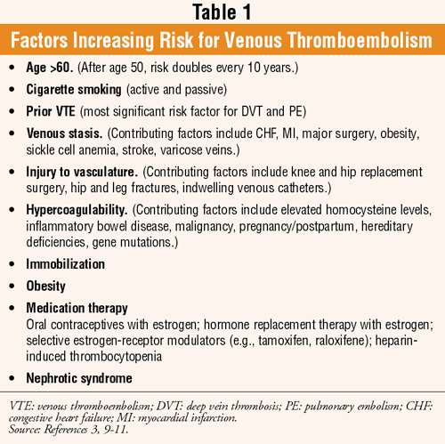
Age is an independent risk factor for thrombotic disease (TABLE 1).1 The majority of venous thrombi occur in either the superficial or deep veins of the leg.2 A deep vein thrombosis (DVT) is stationary clotting blood adhered to the deep vein of the pelvis or an extremity and usually occurs in the calf or thigh.2,3 When the thrombus detaches and thus becomes an intravascular clot, it is referred to as an embolus.4 Venous thromboembolism (VTE) denotes an obstruction arising from the formation of a clot in the venous circulation (i.e., DVT) carried by the blood from the site of origin to plug another vessel (e.g., pulmonary embolism [PE]).5,6

The risks for venous thrombosis are considered immediate (trauma, surgery, temporary immobility); short term (malignancy, pregnancy, severe illness); and long term (multiple prior episodes of thrombosis, acquired and congenital thrombophilic disorders).1 While prognosis is generally good with prompt and appropriate treatment, a long-term complication may ensue--venous insufficiency with or without postphlebitic syndrome.3 Furthermore, a fraction of the patients who present with idiopathic DVT (10%) and recurrent idiopathic DVT (20%) eventually develop clinically overt cancer.1
Pathogenesis
Inappropriate thrombus formation is a disruption of homeostasis and may result from an alteration in any of the factors listed below. The dominant influence, and the one factor that by itself can lead to thrombosis, is endothelial injury.2,5,6
Endothelial Injury: Endothelial injury causes subendothelial collagen exposure and platelet adherence, among other changes; many factors can contribute to the injury, including hypertension, vasculitis, scarred valves, bacterial endotoxins, cholesterolemia, and chemicals from cigarette smoke.2,5,7
Abnormal Blood Flow: Stasis and turbulence are alterations in normal blood flow, which can cause endothelial injury.2 Aneurisms (aortic and arterial dilations) cause local turbulence; a dilated atrium in the presence of atrial fibrillation is a site of significant stasis that may provoke thrombus development.2 Additional conditions are associated with stasis, such as sickle cell anemia and polycythemia, both of which may predispose a patient to thrombosis.2
Hypercoagulability: Hypercoagulability can be described as any alteration of the coagulation pathway that places the patient at risk for thrombosis.2 There are two types of hypercoagulability states; primary disorders are genetically inherited (e.g., mutations in factor V, allelic variations in prothrombin levels) and secondary disorders are acquired (e.g., tissue damage such as surgery, fractures, burns; myocardial infarction; cancer; heparin-induced thrombocytopenia [HIT]).2 Hypercoagulability associated with advancing age is thought to be due to an increase in platelet aggregation and a reduction in prostacyclin, a potent vasodilator and inhibitor of platelet aggregation that is released by the endothelium.2
Symptoms, Signs, and Diagnosis of DVT
Many patients with DVT, and probably most, are asymptomatic.5 There is a risk of sudden death secondary to PE. Complaints of leg swelling, pain, or warmth may be reported; since symptoms are nonspecific, objective testing needs to be performed to establish a diagnosis.5 Upon examination, pain in the back of the knee may be elicited when the foot of the affected leg is dorsiflexed.5 Also in the affected leg, dilation of the superficial veins may be evident and a palpable cord may be felt.
In combination with a meticulous clinical assessment, the noninvasive duplex ultrasonography is the most commonly used test to diagnose DVT.5 The ultrasonography visualizes the venous lining while the Doppler flow study portion of the test detects impaired venous flow (i.e., by measuring rate and direction).3,5 While the test is very sensitive and specific for femoral and popliteal vein thrombosis, small thrombi located in distal veins (e.g., calf) may not be detected using this method.5
Venography, considered the gold standard for the detection of DVT, is expensive and invasive, and requires parenteral radiopaque contrast medium, which may cause anaphylaxis and nephrotoxicity.5 Due to these risks, it is rarely used; venography may be indicated when the ultrasonography is normal but there is a strong suspicion for DVT.3,5 Serum levels of D-dimer (produced when thrombin is generated) are usually elevated, and the patient may have an elevated erythrocyte sedimentation rate and white blood cell count.5
Outcome of the Venous Thrombus
In general, there are four outcomes for a thrombus: 1) propagation--additional platelets and fibrin accumulate, leading to obstruction of the vessel; 2) embolization--thrombi dislodge and travel to other vasculature sites; 3) dissolution--fibrinolytic activity removes thrombi; and 4) organization and recanalization--re-
Embolism
Approximately 99% of all emboli are a part of a thrombus that has been dislodged and is therefore referred to as a thromboembolism.2 Rare types of emboli consist of fat droplets, cholesterol (i.e., atherosclerotic debris), bubbles of air or nitrogen, fragments of tumors, bone marrow, or foreign bodies (e.g., bullets).2 When the term embolism is used, it is understood to mean thrombotic in nature unless otherwise specified.2 A thromboembolic event occurs when an embolus lodges in a vessel, rendering it unable to pass and causing partial or complete occlusion; if necrosis of the distal tissue results, it is referred to as infarction.2 An embolus will lodge in either the pulmonary or systemic circulation (depending on its site of origin), and the clinical outcome will depend on its resting place.2 PE will be discussed below; the interested reader is referred to Reference 2 for a full discussion of systemic emboli.
Clinical Signs and Symptoms of PE: A DVT is the cause of PE in more than 95% of cases.2,5 Approximately 60% to 80% of pulmonary emboli are clinically silent. Sudden death may occur prior to the initiation of therapy.5 The patient may report cough, chest pain, chest tightness, shortness of breath, palpitation, or coughing up blood; dizziness or lightheadedness may occur with a massive PE.5 Symptoms may be confused with those of myocardial infarction or pneumonia.5 The patient may present with tachypnea, tachycardia, and diaphoresis.5 Cyanosis and hypotension may indicate a massive PE, in which case the pulse oximetry or arterial blood gas will show hypoxia and circulatory shock and death may ensue.5 While beyond the scope of this article, the laboratory and diagnostic testing for PE may be found in References 3 and 5.
Treatment Summary for DVT
Treatment of DVT is focused on the prevention of PE; secondarily, it targets relief of symptoms and the prevention of chronic venous insufficiency and postphlebitic syndrome.3 Rapid establishment of anticoagulation is the philosophy of treatment since inadequate anticoagulation in 24 hours increases the risk of PE.1,3 Anticoagulants are administered to all DVT patients; initial treatment consists of an injection of either unfractionated heparin (UFH) or low molecular weight heparin (LMWH; e.g., enoxaparin, dalteparin, tinzaparin) followed by warfarin within 24 to 48 hours.3
Treatment with UFH or LMWH is continued until the patient has achieved full anticoagulation with warfarin (i.e., until the international normalized ratio [INR] is 2.0 to 3.0).3,8,9 Pain control with analgesics (i.e., agents excluding aspirin and other nonsteroidal anti-inflammatory drugs) may be necessary, and parenteral administration may be required for severe symptoms; elevation of legs during periods of inactivity is also a recommended supportive measure.3
Patients over 60 years old who receive UFH may obtain higher serum levels and clinical response (longer activated partial thromboplastin times [aPTT]) as compared to younger patients who receive similar dosages; lower dosages may be required in seniors.8 Complications associated with heparin include bleeding, thrombocytopenia (rare with the LMWHs), urticaria, and although rarely, thrombosis and, anaphylaxis.3 Monitoring parameters include PTT, platelets, hemoglobin, hematocrit, and signs of bleeding.8 Heparin should be discontinued if hemorrhage occurs; severe hemorrhage or overdosage may require treatment with protamine sulfate.8
LMWHs may be used to treat acute DVT on an outpatient basis unless PE is suspected.3
For reducing DVT recurrence, thrombosis extension, and risk of death due to PE, LMWHs are as effective as UFH.3 Since responses are predictable, there is no clear relationship between bleeding and LMWH overdose, and LMWHs do not significantly prolong the aPTT, PTT monitoring is unnecessary.3 Other monitoring parameters include periodic complete blood count including platelets, occult blood, and anti-Xa activity, if available.8 Patients with renal failure are best treated with UFH.3
Warfarin is indicated in the prevention and treatment of VTE and is the drug of choice for long-term anticoagulation for all patients (except in pregnancy and in those with new or worsening VTE during warfarin treatment) due to its low cost and ease of administration.3-5 With a narrow therapeutic index, warfarin predisposes patients to clinically significant drug, food, and herbal interactions (see Reference 5 online), and has the propensity to cause hemorrhage; continuous patient monitoring and education are required to achieve optimal outcomes.5 The therapeutic goal for warfarin therapy in VTE is an INR of 2.0 to 3.0; anticoagulation may be reversed with vitamin K.3,8
Fondaparinux (a selective inhibitor of factor Xa) is indicated for the treatment of DVT and PE, and the prevention of VTE following orthopedic surgery; it should be used with caution in the elderly.5,8 Direct thrombin inhibitors (e.g., lepirudin, desirudin, bivalirudin, argatroban, ximelagatran) are very potent anticoagulants and may be used for the prophylaxis and treatment of VTE and HIT).5
For a complete overview of heparin and other anticoagulation options, dosing in thromboembolic disease, DVT/PE prevention in surgical patients, and specific warnings/precautions, refer to Reference 5 online and individual package inserts.3
The most common adverse effect associated with anticoagulant therapy is bleeding. The risk of a major hemorrhage is related to many factors including anticoagulation intensity (e.g., INR >4.0), history of gastrointestinal bleeding, risk of fall/trauma, recent surgery, and age (i.e., >65 years).5 The Institute for Safe Medication Practices lists heparin among other drugs that have a heightened risk of causing significant harm when used in error.8
Conclusion
A variety of conditions may predispose a senior to thrombosis. Through an understanding of the risk factors for venous thromboembolism and the clinical symptoms and signs, pharmacists may contribute to patient care by referring patients for assessment and evaluation in addition to providing medication counseling and monitoring.
REFERENCES
1. Matulis M, Knovich M, Owen J. Thrombotic and hemorrhagic disorders. In: Hazzard WR, Blass JP, Halter JB, et al, eds. Principles of Geriatric Medicine and Gerontology. 5th ed. New York, NY: McGraw-Hill Inc; 2003:803-817.
2. Mitchell RN, Cotran RS. Hemodynamic disorders, thrombosis, and shock. In: Robbins Pathologic Basis of Disease. 6th ed. Philadelphia, PA: W. B. Saunders Company; 1999:113-138.
3. Beers MH, Porter RS, Jones TV, et al. The Merck Manual of Diagnosis and Therapy. 18th ed. Whitehouse Station, NJ: Merck Research Laboratories.
4. Howland RD, Mycek MJ. Pharmacology. 3rd ed. Philadelphia, PA: Lippincott Williams & Wilkins; 2006:227-244.
5. Haines ST, Zeolla M, Witt DM. Venous thromboembolism. In: DiPiro JT, Talbert RL, Yee GC, et al, eds. Pharmacotherapy: A Pathophysiologic Approach. 6th ed. New York, NY: McGraw-Hill Inc; 2005:373-413.
6. Dorland's Pocket Medical Dictionary. 28th ed. Elsevier Saunders; 2009.
7. Haines ST, Bussey HI. Thrombosis and the pharmacology of antithrombotic agents. Ann Pharmacother. 1995;29:892-905.
8. Semla TP, Beizer JL, Higbee MD. Geriatric Dosage Handbook. 14th ed. Hudson, OH: Lexi-Comp, Inc; 2009.
9. Geerts WH, Pineo GF, Heit JA, et al. Prevention of venous thromboembolism: the Seventh ACCP Conference on Antithrombotic and Thrombolytic Therapy. Chest. 2004;126:338S-400S.
10. Thomas RHMD. Hypercoagulability syndromes. Arch Intern Med. 2001;161:2433-2439.
11. Federman DG, Kirsner RS. An update on hypercoagulable disorders. Arch Intern Med. 2001;161:1051-1056.
To comment on this article, contact
rdavidson@jobson.com.


