US Pharm. 2015;40(4):35-39.
ABSTRACT: Cutaneous fungal infections are commonly caused by dermatophytes. The prevalent dermatophytic infections in the United States include tinea pedis, tinea corporis, tinea cruris, tinea capitis, and tinea unguium. Persons most susceptible to fungal skin infections include immunodeficient or immunosuppressed patients, obese individuals, patients with impaired circulation, and those who are exposed to prolonged moisture or have poor hygiene. Tinea pedis, tinea corporis, and tinea cruris typically are treated topically, unless the infection is extensive, severe, or recalcitrant. Tinea unguium responds best to oral therapy, and tinea capitis must be treated with oral antifungal therapy, since topical agents cannot penetrate the hair shaft. Treatment may last for several weeks to months, making patient adherence an important factor in therapy selection.
Cutaneous fungal infections are superficial infections typically involving the skin, hair, and nails.1 Most commonly, these fungal infections are caused by dermatophytes, but they can also be caused by nondermatophyte fungi and yeast (Candida species).1-4 The term dermatophyte refers to a fungal organism that causes tinea, a fungal infection.5 Thus, dermatophytoses are known as tinea infections, which are further classified by the region of the body infected (e.g., tinea pedis, tinea capitis).1-4
Dermatophytoses are limited to the stratum corneum, nails, and hair shafts because they require keratin for growth.3,4,6 The prevalent dermatophytic infections in the United States include tinea pedis (foot), tinea corporis (body), tinea cruris (groin), tinea capitis (scalp), and tinea unguium (nail). In the U.S., there are three dermatophyte genera that cause infections: Microsporum, Epidermophyton, and Trichophyton. Trichophyton is the most prevalent genus, accounting for approximately 80% of dermatophytic infections in the U.S.4,6 The most common mode of transmission of dermatophytes is by direct contact with other people (anthropophilic organisms), but transmission also occurs via contact with animals (zoophilic organisms), the soil (geophilic organisms), and fomites.4 Individuals most susceptible to fungal skin infections include those who are obese, immunodeficient, or immunosuppressed or have impaired circulation.1 Fungal infections are also more likely to occur with prolonged exposure to sweaty clothes or bedding, poor hygiene, and residence in warm, humid climates.1,2
The classic appearance of a cutaneous tinea infection is a central clearing surrounded by an active border of redness and scaling, which gives rise to the more common name, ringworm.2,4 One key point in recognizing a cutaneous fungal infection is the location; tinea infections have no mucosal involvement, since dermatophytes invade only keratinized tissue. 4 Despite having a classic appearance, tinea infections may be similar in appearance to many other dermatologic conditions and are often misdiagnosed and, therefore, mistreated.2 This article will guide pharmacists to recognize the most common fungal infections, understand the most effective treatment options, and provide counseling for treatment and prevention.
Types of Dermatophytoses
Tinea Pedis: Tinea pedis is the most prevalent cutaneous fungal infection.1,2 Frequently referred to as athlete’s foot, it affects approximately 26.5 million Americans per year.1 It is estimated that approximately 70% of people will have tinea pedis during their lifetime.
Four clinically accepted variants of tinea pedis exist; however, they sometimes overlap. The most common variant is intertriginous, which is characterized by fissuring, scaling, or maceration of the interdigital areas; foul odor; itching; and a stinging sensation.1 The infection often involves the lateral toe webs and may spread to the sole or instep of the foot. Warm, humid conditions may aggravate the area. The second variant is a chronic papulosquamous type that often occurs on both feet. Mild inflammation and dispersed scaling of the skin on the soles of the feet are characteristic of this type. The third variant is composed of small vesicles or vesicopustules on the instep and plantar surface. Scaling of the skin in this area, as well as the toe webs, is observed. The fourth variant involves macerated, denuded, weeping ulcerations on the sole of the foot. Odor is common with this type. This variant is often complicated by opportunistic gram-negative bacteria. Differential diagnosis includes eczema, contact dermatitis, psoriasis, and pitted keratolysis.2
Adults typically have an increased risk of tinea pedis compared with children, owing to increased exposure opportunities.1 Persons who use public pools or bathing facilities are at increased risk.1-3,7 Individuals who participate in high-impact activities that cause chronic trauma to the foot and those who wear occlusive footwear are also at increased risk.1,2 Treatment with topical agents is the preferred therapy.5,7 Systemic antifungal agents may be required for failed treatment with topical agents, extensive disease, or an immunocompromised state.5
Tinea Corporis: Also called ringworm, tinea corporis may present in multiple ways and on multiple areas of the body.1,2 Lesions frequently manifest as small, circular, erythematous, scaly spaces.1-3,5 Central clearing occurs as the borders spread and vesicles or pustules develop. Tinea corporis may occur on any body part, depending upon the type of dermatophyte infection. Zoophilic dermatophytes frequently infect areas of exposed skin, whereas anthropophilic dermatophytes infect occluded areas or sites of trauma. Differential diagnosis includes eczema, psoriasis, and seborrheic dermatitis.5
Tinea Cruris: Tinea cruris, or jock itch, occurs on the medial and upper area of the thighs and groin area and is more common in males than in females.1 The scrotum itself often is not affected.5,8 Signs of excessive moisture, pruritus, and burning are often present.2 Risk factors for tinea cruris include infection with tinea pedis, obesity, diabetes, and immunodeficiency. Differential diagnosis includes candidiasis, intertrigo, erythrasma, psoriasis, and seborrheic dermatitis.2
Tinea Capitis: Tinea capitis is also called ringworm of the scalp. The incidence of this form is not known; however, it occurs most frequently in children exposed through contact with other children or pets.2,3 Three types of tinea capitis exist: black dot, gray patch, and favus.5,8 Trichophyton tonsurans frequently causes black dot tinea capitis and is the predominant variant observed in the U.S.2,5,8 Gray patch tinea capitis occurs in epidemic and endemic forms; however, the epidemic form is no longer documented in the U.S. The endemic form, which is caused by Microsporum canis, is often spread by cats and dogs. Favus, which rarely occurs in the U.S., is characterized by spores, air spaces, and fragmented hyphae, and occurs more frequently in Eastern Europe and Asia.9
Black dot tinea capitis is often asymptomatic initially. An erythematous, scaling patch on the scalp enlarges over time, and alopecia occurs.3 Hairs within the patches break, and a black dot (caused by detritus within the follicular opening) appears.5,10 If black dot tinea capitis is left untreated, the alopecia and scarring may be permanent.3,10 On occasion, the lesion may change and become elevated, tender, highly inflamed nodules known as kerion. Kerion formation is due to an immune response to the fungus. Lymphadenopathy may occur with kerion. Gray patch tinea capitis presents as circular patches of alopecia with prominent scaling.10 Kerion formation may occur with gray patch tinea capitis infection.
Tinea capitis must be treated with systemic antifungal agents, since topicals cannot penetrate the hair shaft.5,10 Adjunctive treatment with antifungal shampoos may be recommended. Asymptomatic carriers of dermatophytes may be a source of reinfection. Sharing of fomites such as hats, combs, and brushes should be avoided.5 Differential diagnosis includes alopecia areata, atopic dermatitis, bacterial infection, psoriasis, and seborrheic dermatitis.5
Tinea Unguium: This disorder, also known as onychomycosis, is caused most frequently by dermatophytes, but nondermatophytes and Candida species also can cause it.7 Annually, more than 2.5 million people in the U.S. are treated for tinea unguium. Affected nails often become thick, rough, yellow, opaque, and brittle.1,2,5 The nail may separate from the nail bed, and the dermis surrounding the infected nail may be hyperkeratotic.7 Risk factors include diabetes, trauma, family history, tinea pedis, smoking, extended periods of water exposure, and immunodeficiency. Differential diagnosis includes psoriasis, eczema, lichen planus, and trauma.2,5 Treatment requires oral therapy for an extended period, at least 6 to 12 weeks, depending upon the location of the infection.5,7 Failure rates with oral therapy typically are high, and topical treatment generally is not effective.
Tinea Incognito: Tinea incognito is a dermatophyte infection that is modified because of treatment with a corticosteroid.2,3 Margins may be lost, and the area may be more widespread. Tinea incognito requires a thorough patient history and should be considered when a corticosteroid has been used to treat a rash that appeared to have cleared, but returned unresolved.
Candida Yeast Infections
Candida is part of normal body flora, but it is also a common cause of yeast infections.3 When the normal balance of flora is disturbed, an acute infection may occur. Risk factors include antibiotics, corticosteroids, diabetes, obesity, immunosuppression, and immunodeficiency.2,3 Additionally, Candida thrives in warm, moist conditions.2 An infection often presents as red lesions with accompanying satellite papules and pustules. Common areas of infection are the mouth and genital region. Differential diagnosis includes tinea corporis.
Treatment of Fungal Skin Infections
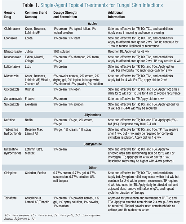
Topical Therapy: Tinea pedis, tinea corporis, and tinea cruris generally respond well to topical therapy.1 Many of these treatments are available as nonprescription formulations. Commonly used topical therapies are described in TABLE 1.1,11 Topical agents are available as ointments, creams, powders, and aerosols, and are well-tolerated overall. Rare cases of mild skin irritation, burning, itching, or dryness have been reported.1 Drug-drug interactions are unlikely with topical therapy.

Multiple combination products incorporating an antifungal plus a corticosteroid are available. Combination therapy with antifungals and corticosteroids is not currently recommended in clinical guidelines.12 Clinical cure rates have been demonstrated for combination therapy; however, the quality of the studies was poor owing to imprecision and bias, and relapse rates could not be assessed.
Patient adherence may be affected by the product chosen. Therefore, the selection of a drug or product should be made based on the patient’s daily habits and activities, as well as patient-specific characteristics such as concomitant disease states, age, and drug sensitivities.
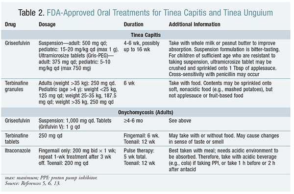
Oral Therapy: Oral therapy can be recommended for the treatment of tinea pedis, tinea corporis, and tinea cruris if the infection is extensive, severe, or recalcitrant.6 See TABLE 2. However, tinea capitis must be treated with oral antifungal therapy, since topical agents do not penetrate the hair shaft, and tinea unguium responds better to oral therapy than to topical treatment.5,6

There are currently two FDA-approved pediatric treatment options for tinea capitis: griseofulvin and terbinafine.5,6,13 Griseofulvin is available as a suspension and as ultramicrosize tablets, but the tablets may be preferred, given the bitter taste of the suspension.13 For griseofulvin, there is some discrepancy regarding treatment duration for tinea capitis. Manufacturer labeling recommends 4 to 6 weeks, but other sources advise 6 to 12 weeks, and possibly up to 16 weeks.13 The American Academy of Pediatrics recommends that griseofulvin be continued for 2 weeks after clinical resolution of the infection.14 In 2007, terbinafine was approved to treat tinea capitis in patients aged 4 years and older.13 Terbinafine is dosed according to weight and is given once daily for 6 weeks. Off-label uses of itraconazole syrup (5 mg/kg for 4 weeks) and fluconazole (6 mg/kg daily for 3-6 weeks or 6 mg/kg once weekly) are alternative therapies.3,5 Because of increasing resistance to griseofulvin, alternative regimens may be preferred for the treatment of tinea capitis; however, griseofulvin remains the drug of choice for kerion and when the etiologic agent is a Microsporum species.5
There are several different oral treatment approaches for onychomycosis. Oral griseofulvin, terbinafine, itraconazole, or fluconazole may be useful, but dosages and treatment duration vary according to the location of the infection (e.g., toenails or fingernails).6 These antifungal therapies may be effective only if the onychomycosis is caused by dermatophytes; if Candida is the etiologic agent, the infection may be resistant to oral antifungal therapy. Fluconazole is not approved for the treatment of onychomycosis, but pulse dosing may be used off-label (150-300 mg once a week for 3-6 months for fingernails or 6-12 months for toenails). Some researchers believe that oral antifungal therapy should be continued past the recommended treatment duration, at least until the infected nail is replaced by normal growth; however, this may take up to 9 to 12 months.5,6
The use of the oral antifungal agents is not without side effects or significant drug interactions.13 Common adverse effects of griseofulvin include rash, headache, nausea and vomiting, and photosensitivity; additionally, long-term use of griseofulvin may result in hepatotoxicity. Notable drug interactions with griseofulvin include barbiturates, alcohol, cyclosporine, oral contraceptives, aspirin, and warfarin. Adverse effects of itraconazole include diarrhea, rhinitis, dyspepsia, pruritus, and hypertension. Notable drug interactions with itraconazole include terfenadine, astemizole, diazepam, oral triazolam, oral midazolam, cisapride, and hydroxymethyl glutaryl coenzyme A reductase inhibitors. Fluconazole may cause nausea and vomiting, rash, abdominal pain, and changes in taste. Like itraconazole, fluconazole has many drug interactions and should also be avoided in patients with renal impairment or hepatic disease. All oral antifungals require routine liver function tests.
Nonpharmacologic Therapy: Good skin care, including regular bathing and complete drying of the skin, is essential for preventing fungal skin infections. Prolonged exposure of the affected area(s) to moisture should be avoided. To prevent the recurrence of tinea pedis, walking barefoot in areas such as public bathrooms, locker rooms, and showers should be avoided. Affected individuals should also consider nonocclusive shoes, absorbent socks, and powder to control the moisture content.
When tinea capitis is confirmed, all contaminated combs, brushes, hats, and bedding should be cleaned. Diagnosed children may return to school once treatment of tinea capitis has begun; however, the sharing of grooming utensils, hats, and bedding should be avoided for at least 14 days.5
Conclusion
Proper identification and treatment of fungal skin infections remains a growing health concern. Pharmacists should refer patients with suspected tinea infections to their primary care provider for diagnosis confirmation, and then work in collaboration to effectively manage the infection with pharmacologic and nonpharmacologic treatment recommendations. Pharmacists are well positioned to encourage proper use of and adherence to lengthy treatment regimens. Since patient adherence may be affected by product selection, the pharmacist should consider patient characteristics and lifestyle to ensure that the appropriate product and formulation is chosen.
REFERENCES
1. Newton GD, Popovich NG. Fungal skin infections. In: Krinsky DL, Berardi RR, Ferreri SP, et al, eds. Handbook of Nonprescription Drugs. 17th ed. Washington DC: American Pharmacists Association; 2012:757-771.
2. Goldstein AO, Smith KM, Ives TJ, Goldstein B. Mycotic infections. Effective management of conditions involving the skin, hair, and nails. Geriatrics. 2000;55(5):40-52,45-47,51-52.
3. Robinson J. Fungal skin infections in children. Nurs Stand. 2012;27:52-54,56,58.
4. Hainer BL. Dermatophyte infections. Am Fam Physician. 2003;67:101-108.
5. Ely JW, Rosenfeld S, Seabury Stone M. Diagnosis and management of tinea infections. Am Fam Physician. 2014;90:702-710.
6. Vander Straten MR, Hossain MA, Ghannoum MA. Cutaneous infections: dermatophytosis, onychomycosis, and tinea versicolor. Infect Dis Clin North Am. 2003;17:87-112.
7. Flint WW, Cain JD. Nail and skin disorders of the foot. Med Clin North Am. 2014;98:213-225.
8. Hawkins DM, Smidt AC. Superficial fungal infections in children. Pediatr Clin North Am. 2014;61:443-455.
9. Drake LA, Dinehart SM, Farmer ER, et al. Guidelines of care for superficial mycotic infections of the skin: tinea capitis and tinea barbae. J Am Acad Dermatol. 1996;34:290-294.
10. Fuller LC, Child FJ, Midgley G, Higgins EM. Diagnosis and management of scalp ringworm. BMJ. 2003;326:539-541.
11. Clinical Pharmacology [online database]. Tampa, FL: Elsevier/Gold Standard; 2014.
12. El-Gohary M, van Zuuren EJ, Fedorowicz Z, et al. Topical antifungal treatments for tinea cruris and tinea corporis. Cochrane Database Syst Rev. 2014;(8):CD009992.
13. Lexicomp Online [online database]. Hudson, OH: Wolters Kluwer Health; 2015.
14. Tinea capitis. In: Pickering LK, Baker CJ, Kimberlin DW, Long SS, eds. Red Book: 2009 Report of the Committee on Infectious Diseases. 28th ed. Elk Grove Village, IL: American Academy of Pediatrics; 2009:662.
To comment on this article, contact rdavidson@uspharmacist.com.


