US Pharm. 2020;12(45):HS12-HS16.
ABSTRACT: Sigmoid volvulus (SV), the most frequent type of colonic volvulus, is a common cause of bowel obstruction. Although SV occurs in the setting of constipation and congenitally elongated colon, among other predisposing conditions, the precipitating factor in SV formation has not been confirmed. Most cases of SV occur in debilitated elderly patients with multiple comorbidities who may not be candidates for surgical resection. Plain abdominal radiography is used to diagnose SV in most cases, with computed tomography and laparoscopy used as confirmatory tests when necessary. Owing to the high risk of recurrence, SV is typically managed via endoscopic detorsion and decompression, followed by nonemergent surgical resection.
Volvulus, a condition in which the intestine twists upon itself and its mesentery and causes obstruction, involves the colon or small bowel.1 Small-bowel volvulus is more likely in children, whereas colonic volvulus (CV) occurs more often in adults.2,3 In the United States, CV is the third most common cause of large-bowel obstruction and is responsible for approximately 5% of all cases of intestinal obstruction and 10% to 15% of all cases of large-bowel obstruction. The sigmoid colon is the most common site of large-bowel torsion (80%), followed by the cecum (15%), transverse colon (3%), and splenic flexure (2%). Prompt diagnosis and treatment are critical in order to avoid the development of bowel ischemia.2
Sigmoid volvulus (SV), the most common type of CV, is a frequent cause of bowel obstruction. Although SV occurs in the setting of constipation and congenitally elongated colon, among other predisposing conditions, the precipitating factor in SV formation has not been confirmed.1 Owing to the high risk of recurrence, SV is typically managed via endoscopic detorsion and decompression followed by nonemergent surgical resection.1,4
Epidemiology and Etiopathogenesis
SV accounts for 2% to 5% of colonic obstructions in Western countries and 20% to 50% of such obstructions in Eastern countries.2 African, Asian, Middle Eastern, South American, Eastern European, and Northern European countries, as well as Turkey, are endemic regions for SV. The community-based incidence of SV in the U.S. is 1.67 per 100,000 persons per year. SV typically affects adults, and the highest incidence is in the fourth through eighth decades of life. The disorder is more common in males, and reported ratios range from 2:1 to 10:1. SV accounts for 4% of all intestinal volvulus cases in children and is usually accompanied by congenital anomalies. Nearly one-third of elderly patients have a history of SV attack, and 50% to 85% have serious coexisting diseases.5
SV occurs most frequently in elderly, institutionalized, and chronically constipated persons, and it is the most common cause of large-bowel obstruction during pregnancy. In the U.S., the factors that predispose to volvulus in adults are largely acquired. The acquired causes of SV mainly include conditions that cause an increase in the span of the sigmoid colon, such as chronic constipation, infections, neuropsychiatric disorders, and electrolyte abnormalities. Intra-abdominal adhesions, which can be acquired secondary to surgery, injury, and infections, make torsions more likely. Hirschsprung disease (congenital absence of ganglions in the bowel segment, most commonly in the colon) causes dilatation and elongation of the proximal bowel, thus increasing the chance of volvulus. Psychotropic drugs interfere with colonic motility and are etiologically implicated in the high incidence seen in patients in psychiatric facilities. Other etiologic factors have been noted in different regions of the world. The high incidence of SV in areas such as Russia, India, Iran, Norway, and Africa has been attributed to the high-fiber vegetable diets prevalent in these regions. This type of diet causes increased gas and sigmoid-colon elongation. In Brazil, Chagas disease is the chief cause of megacolon, and 30% of 365 megacolon patients in São Paulo were reported to have SV.6-8
Clinical Features
Usual presenting symptoms for patients with SV include constipation, diarrhea, vomiting, abdominal distention, tense and tender abdomen, bright red blood in the stool, and sluggish gut sounds. On digital rectal examination, the rectum may be empty or may contain fresh blood. SV is an acute surgical emergency because in a very short period of time it increases the likelihood of complications such as gut ischemia, gangrenous bowel segment, peritonitis, shock, sepsis, and perforation.6
SV twists of less than 180° are known as physiological volvulus. Luminal obstruction (obstructive volvulus) and vascular compromise (strangulating volvulus) occur when torsion exceeds 180° and 360°, respectively. The clinical presentation of SV depends more on the rapidity of colonic twisting than on the degree of torsion. TABLE 1 lists the clinical classification of SV.8
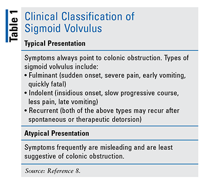
The earlier the presentation of SV, the worse the prognosis will be. This is because fulminant volvulus usually presents early owing to ischemic pain. Nevertheless, iatrogenic delay in diagnosis, which commonly occurs in nursing home residents, worsens the prognosis. The triad of abdominal distention, pain, and constipation, although frequently misconstrued as classical signs of SV, is neither specific nor sensitive. Vomiting, which is usually a late and uncommon symptom of distal colonic obstruction, is frequently reported in SV. Early vomiting, which is a phenomenon of vagal reflex in fulminant volvulus, predicts a poor prognosis. Physical signs are mostly nonspecific and simply indicate distal colonic obstruction. However, emptiness of the left iliac fossa is a pathognomonic sign of SV.8
Diagnosis
The diagnosis of SV is established through clinical and radiologic findings. In the majority of patients, a thorough physical examination and abdominal radiographs are adequate for achieving the diagnosis. Diagnostic imaging often includes confirmatory imaging with a contrast enema or computed tomography (CT) imaging. The use of sigmoidoscopy for therapeutic as well as diagnostic procedures makes this a valuable testing tool in SV. For patients with abdominal emergencies, laparoscopy provides diagnostic accuracy and therapeutic options, avoids extensive preoperative studies, averts delays in operative intervention, and appears to reduce morbidity. A diagnosis of SV is also made via radiologic signs that are mostly named for common patterns or objects readily recognizable in everyday life. The objective behind these associations is to aid in the understanding and diagnosis of the disease process. These signs may be seen in different imaging modalities, such as plain radiograph and CT.9-11
Plain Abdominal Radiography (X-Rays): Plain abdominal radiographs usually show a dilated sigmoid colon and multiple small or large intestinal air-fluid levels. Described diagnostic x-ray signs are the omega or horseshoe sign; bird’s beak sign; inverted V sign; Y sign; northern exposure sign; coffee bean sign; bent inner tube or ace-of-spades sign; left pelvic overlap or left flank overlap sign; liver overlap sign; and empty left iliac fossa sign. Plain abdominal radiography has been found to be diagnostic in 57% to 90% of patients.5
CT Scan: In cases in which clinical assessment and plain abdominal radiographs are insufficient to confirm the diagnosis of CV, contrast enema or CT imaging may be helpful. In general, a water-soluble contrast medium is preferable to barium contrast because the latter could cause chemical peritonitis in the setting of a perforated colon. Contrast-enhanced CT imaging is the preferred confirmatory diagnostic technique for SV because it is noninvasive, easily obtainable, and accurate for SV, in addition to having the advantage of identifying incidental pathology that may be missed with plain radiographs or fluoroscopic contrast studies. In one study, the positive diagnostic yield of CT for SV was found to be 89%. Other conditions whose presentation can mimic that of CV, such as pseudo-obstruction or obstruction caused by a neoplasm, can be differentiated with the above modalities.12 The coffee bean sign, which was identified in 76% of patients in one study, is thought to be specific for the diagnosis of SV and may be the best initial feature suggesting the disorder. CT is often used to assess bowel ischemia, the fundamental complication of SV. Bowel ischemia can progress to infarction, perforation, and death.13
Contrast Enema (Barium): A barium enema may assist with the diagnosis of SV when the noncontrasted study is equivocal. The appearance of a bird’s beak (point of twisted bowel) or ace-of-spades sign is due to narrowing of the rectosigmoid at the neck of the volvulus, causing complete or partial obstruction. A distended and downwardly displaced transverse colon can mimic SV by producing a pseudo-volvulus. Barium has the potential to cause significant complications by forming an impaction, which occludes the lumen of bowel, resulting in constipation or complete obstruction. Inactive and dehydrated elderly patients, as well as neonates, are at greater risk for impaction. This risk can be minimized by copious fluid intake, prompt evacuation of the barium, and use of a stool softener or laxative following the procedure. Perforation due to catheter-tip insertion and overinflation is potentially the most serious complication, occurring in approximately 0.02% to 0.04% of cases. Free barium is inert, but the dyes, bacteria, and partially digested food matter dumped into the peritoneum cause peritonitis, and third-spacing of fluid leads to hypovolemia. Bacteremia has been found in up to 23% of patients following barium enema, and in rare cases it can cause septicemia. Barium can also induce an inflammatory reaction wherein the barium crystals become coated with a fibrin membrane, followed by fibrosis and granuloma formation. Contraindications to contrast-enema evaluation include evidence of colonic perforation (unless used to assess for perforation), ischemic colon, toxic megacolon, hypovolemic shock, peritonitis, and other potentially unstable clinical conditions.14,15
Sigmoidoscopy: This procedure helps establish the diagnosis of SV. The classical finding on sigmoidoscopy is a spiral sphincterlike twist of the lumen, usually 20 cm to 30 cm from the anal verge; additionally, inability to insert the endoscope into the proximally twisted site helps lead to the correct diagnosis. Sigmoidoscopy allows for direct visualization of the bowel mucosa viability and may also be used in the differential diagnosis of SV by identifying the other causes of bowel obstruction, such as bowel malignancies or megacolon. Although sigmoidoscopy is thought to have a high diagnostic value (a 76%-100% diagnostic success rate) in patients with SV, no quantitative data are available that address the overall diagnostic role of sigmoidoscopy. The main complications of sigmoidoscopy-treated SV, as well as the most common causes of sigmoidoscopy-related death, are bowel perforation, peritonitis, shock, fluid-electrolyte imbalances, renal insufficiency, and cardiopulmonary problems.16
Laparoscopy: Laparoscopy is a surgical diagnostic procedure. Emergency laparotomy and resection with or without primary anastomosis are indicated when nonoperative methods fail or when there is evidence of strangulation, infarction, or perforation. Postoperative mortality ranges from 6% to 60%. Factors associated with poor prognosis include advanced age, delayed diagnosis, presence of intestinal infarction, peritonitis, and shock at presentation. Recurrence has been reported in up to 60% of cases for which surgery may be indicated. Approaches for preventing recurrence include endoscopic decompression of the volvulus followed by either resection or sigmoidopexy.17
Prognosis
The most important risk factor for mortality is the delay in sigmoid decompression, which eventually leads to intestinal ischemia and gangrene. Another interesting finding is the relationship between leukocyte count and SV prognosis. It was demonstrated that if the leukocyte count was below 5,000 or up to 20,000/mm3 at the time of admission, the possibility of intestinal gangrene was 12.5% and 100%, respectively. The association between a prognosis of SV and gangrene was found to be significant.18 Other risk factors that contribute to poor prognosis include toxic or septic shock, advanced age, and serious disease affecting the heart, lungs, kidney, and other organs.
Management
The management of SV involves relieving the obstruction and preventing recurrent attacks. Since the introduction of endoscopic detorsion in the 1940s, this approach—along with subsequent resection—has become the primary therapeutic modality. Detorsion can be performed via barium enema, rigid proctoscopy, flexible sigmoidoscopy, or colonoscopy. It has been reported that 24% of sigmoidoscopic approaches will not find the site of torsion; therefore, the use of colonoscopy is encouraged. Overall, decompression has been found to be successful in 70% to 80% of cases. Barium enema (if not previously used diagnostically) may result in detorsion of the volvulus in about 5% of patients. Sigmoidoscopic detorsion without resection results in a high recurrence rate (18%-90%) and a mortality rate of 5% to 14%. In one study, for cases in which endoscopic detorsion was possible, the success rate was associated with absence of abdominal tenderness, laxative use, and history of open abdominal surgery. Care should be taken in the selection of patients for endoscopic detorsion. Patients exhibiting signs and symptoms of sepsis, fever, leukocytosis, and peritonitis should be taken directly to the operating room for exploration. Patients who fail endoscopic decompression, have gangrenous bowel identified on endoscopy, or exhibit signs and symptoms of sepsis should be expeditiously prepared for surgery.19,20
Conclusion
SV remains a cause of potentially lethal intestinal obstruction that is typically seen in debilitated elderly patients with multiple comorbidities who may not be candidates for surgical resection; it also occurs in institutionalized patients. The endoscopic procedure has an important diagnostic and therapeutic role given its effectiveness and safety in resolving SV, despite the high recurrence expected. Elective surgery in these high-risk patients seems to be safe and preferable to emergency procedures, which have high morbidity and mortality, but randomized, controlled studies with larger numbers of subjects are needed in order to evaluate this hypothesis.21
REFERENCES
1. Osiro SB, Cunningham D, Shoja MM, et al. The twisted colon: a review of sigmoid volvulus. Am Surg. 2012;78(3):271-279.
2. Kapadia MR. Volvulus of the small bowel and colon. Clin Colon Rectal Surg. 2017;30(1):40-45.
3. Meytes V, Schulberg SP, Morin N, Glinik G. Undiagnosed hypothyroidism presenting with sigmoid volvulus. J Surg Case Rep. 2016;2016(4):rjw033.
4. Tin K, Sobani ZA, Anyadike N, et al. Percutaneous endoscopic sigmoidopexy using T-fasteners for management of sigmoid volvulus. Int J Colorectal Dis. 2017;32(7):1073-1076.
5. Atamanalp SS. Sigmoid volvulus. Eurasian J Med. 2010;42(3):142-147.
6. Asghar MS, Tauseef A, Shariq H, et al. Sigmoid volvulus: a rare but unique complication of enteric fever. J Community Hosp Intern Med Perspect. 2020;10(1):45-49.
7. Ballantyne GH, Brandner MD, Beart RW Jr, Ilstrup DM. Volvulus of the colon. Incidence and mortality. Ann Surg. 1985;202(1):83-92.
8. Raveenthiran V, Madiba TE, Atamanalp SS, De U. Volvulus of the sigmoid colon. Colorectal Dis. 2010;12(7):e1-e17.
9. Rao KJ, Podili NK, Rajagopal M. Review of sigmoid volvulus: a 5 years experience in tertiary care hospital, Visakhapatnam. J Evid Based Med Healthc. 2018;5(25):1948-1953.
10. Kirshtein B, Roy-Shapira A, Lantsberg L, et al. The use of laparoscopy in abdominal emergencies. Surg Endosc. 2003;17(7):1118-1124.
11. Singh Y, Islam S, Arra A, et al. The steel pan sign of sigmoid volvulus—a case series. Int J Surg Case Rep. 2017;41:332-335.
12. Vogel JD, Feingold DL, Stewart DB, et al. Clinical practice guidelines for colon volvulus and acute colonic pseudo-obstruction. Dis Colon Rectum. 2016;59(7):589-600.
13. Levsky JM, Den EI, DuBrow RA, et al. CT findings of sigmoid volvulus. Am J Roentgenol. 2010;194(1):136-143.
14. Ramakrishnan K, Scheid D. Opening Pandora’s box: the role of contrast enemas in abdominal imaging. Internet J Gastroenterology. 2002;2:1-11.
15. American College of Radiology. ACR-SPR practice parameter for the performance of pediatric fluoroscopic contrast enema examinations. Revised 2016 (resolution 9). www.acr.org/-/media/ACR/Files/Practice-Parameters/FluourConEnema-Ped.pdf. Accessed November 20, 2020.
16. Atamanalp SS, Atamanalp RS. The role of sigmoidoscopy in the diagnosis and treatment of sigmoid volvulus. Pak J Med Sci. 2016;32(1):244-248.
17. Wai CT, Lau G, Khor CJ. Clinics in diagnostic imaging (105): sigmoid volvulus causing intestinal obstruction, with successful endoscopic decompression. Singapore Med J. 2005;46(9):483-487.
18. Maddah G, Kazemzadeh GH, Abdollahi A, et al. Management of sigmoid volvulus: options and prognosis. J Coll Physicians Surg Pak. 2014;24(1):13-17.
19. Gingold D, Murrell Z. Management of colonic volvulus. Clin Colon Rectal Surg. 2012;25(4):236-244.
20. Iida T, Nakagaki S, Satoh S, et al. Clinical outcomes of sigmoid colon volvulus: identification of the factors associated with successful endoscopic detorsion. Intest Res. 2017;15(2):215-220.
21. da Rocha MC, Capela T, Silva MJ, et al. Endoscopic management of sigmoid volvulus in a debilitated population: what relevance? GE Port J Gastroenterol. 2020;27(3):160-165.
The content contained in this article is for informational purposes only. The content is not intended to be a substitute for professional advice. Reliance on any information provided in this article is solely at your own risk.
To comment on this article, contact rdavidson@uspharmacist.com.


